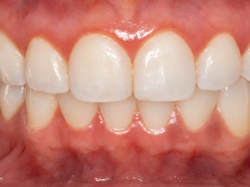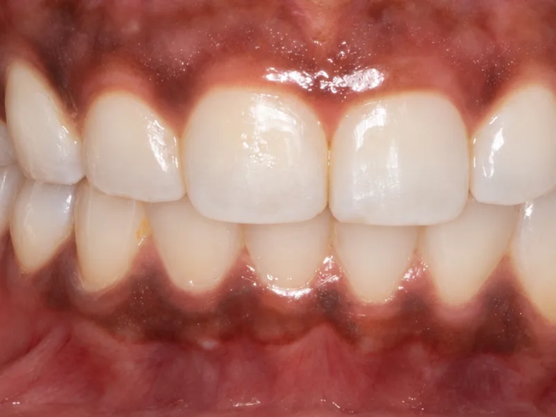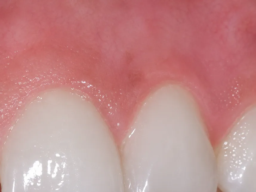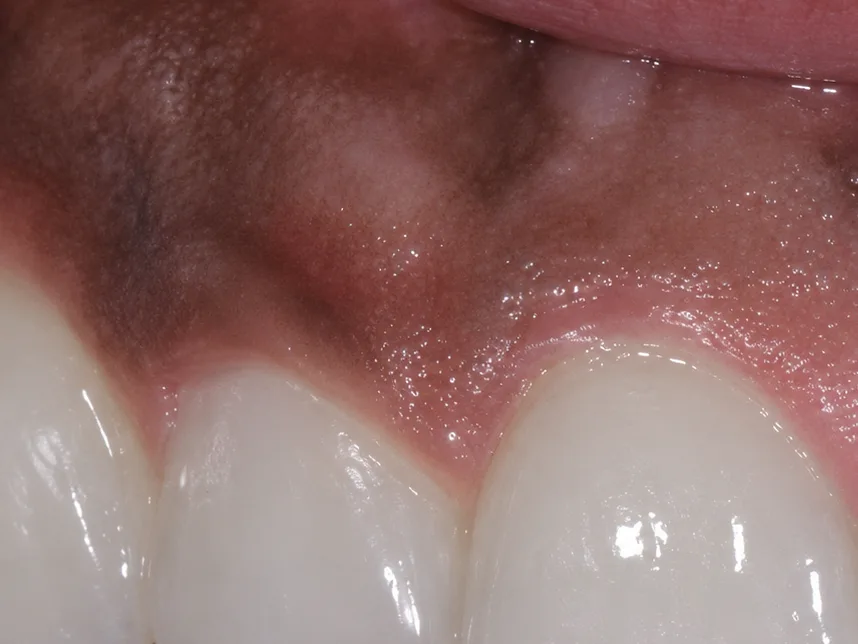
What Causes Dark Gums?
Gum colour varies naturally between individuals. Brown or darker areas commonly reflect melanin, the pigment also responsible for skin colour.
Other possible causes include smoking, certain medications, previous inflammation and deposits from dental materials. Some pigmentation can be associated with medical conditions, so a diagnosis should come before cosmetic treatment.
A new, changing, irregular or unexplained dark patch should be examined before any attempt to remove it. Further investigation or a biopsy may be necessary if the diagnosis is uncertain.
What Is Gum Depigmentation?
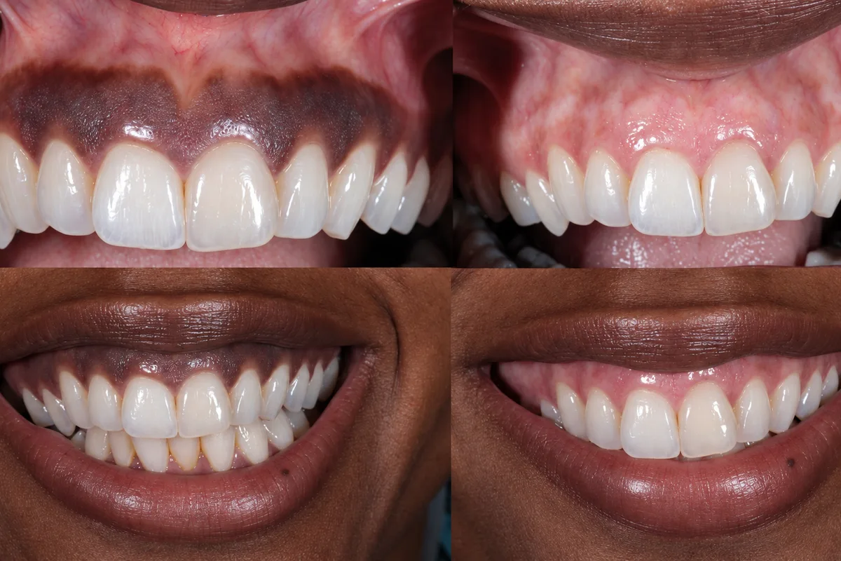
Also called gingival depigmentation, treatment removes or treats selected superficial pigmented tissue, allowing the surface to heal.
The aim is to reduce visible pigmentation while preserving healthy gum tissue. It does not whiten the teeth, treat gum disease or necessarily change the shape of the gum line. The final colour depends on your natural tissues and healing response.
Who May Be Suitable?
Treatment may be considered when:
- The pigmentation has been assessed and identified as suitable for cosmetic treatment.
- The gums are healthy, or existing inflammation has been treated.
- The patient understands the expected result and possibility of recurrence.
- Medical history and healing factors support the proposed procedure.
Not every dark area responds to the same approach. Addressing an underlying cause may be more appropriate than removing pigment.
Types of Gum Depigmentation
Gum depigmentation can be performed using several techniques. The choice depends on the cause and extent of pigmentation, gum thickness, medical history and the clinician’s assessment.
Naturally pigmented gums can be healthy. Treatment is cosmetic once the pigmentation has been assessed. New, changing or unexplained dark areas require diagnosis before treatment.
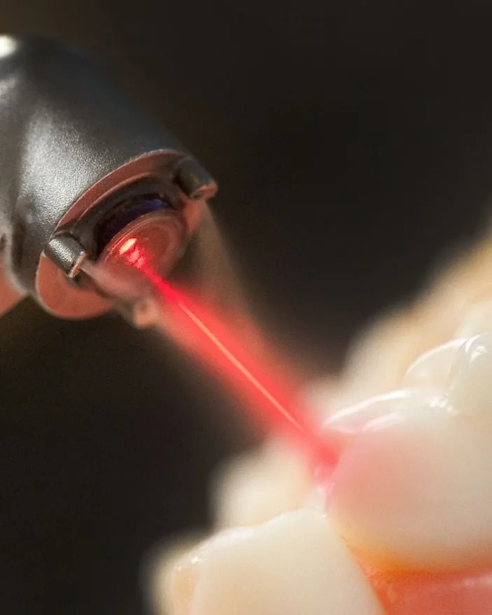
Laser Depigmentation
A suitable dental laser treats the superficial tissue containing melanin pigment. Depending on the laser used, it can provide good bleeding control during the procedure.
Discomfort, healing time and results vary. Laser treatment does not guarantee painless treatment or prevent pigmentation from returning.
Surgical Blade Depigmentation
Also called surgical stripping, this technique uses a scalpel to carefully remove the superficial pigmented tissue. The area then heals, and a protective dressing may be placed where appropriate.
It is an established approach that does not require laser equipment. Bleeding and postoperative tenderness are managed through appropriate clinical care.
Rotary Abrasion or Ceramic Bur Depigmentation
A suitable rotary instrument removes selected superficial pigmented tissue. Careful control of pressure and depth is essential to protect the gum margins and underlying structures.
Electrosurgical Depigmentation
An electrosurgical instrument uses electrical energy to remove superficial tissue while helping control bleeding. Precise application is needed to avoid excessive heat damage to neighbouring tissues.
Cryosurgical Depigmentation
Controlled freezing treats the pigmented tissue, which subsequently sheds as the area heals. Treatment depth and the surrounding tissues require careful management. Availability varies between clinics.
| Technique | How It Works | Key Consideration |
|---|---|---|
| Laser | Treats superficial pigmented tissue using laser energy | Bleeding control varies by laser; heat must be carefully controlled |
| Surgical blade | Removes superficial tissue with a scalpel | Requires bleeding management and wound care |
| Rotary abrasion / ceramic bur | Mechanically removes the pigmented surface | Depth and pressure require precise control |
| Electrosurgery | Uses electrical energy to remove tissue | Thermal injury must be avoided |
| Cryosurgery | Uses controlled freezing | Treatment depth and healing response can vary |
Laser
- How It Works
- Treats superficial pigmented tissue using laser energy
- Key Consideration
- Bleeding control varies by laser; heat must be carefully controlled
Surgical blade
- How It Works
- Removes superficial tissue with a scalpel
- Key Consideration
- Requires bleeding management and wound care
Rotary abrasion / ceramic bur
- How It Works
- Mechanically removes the pigmented surface
- Key Consideration
- Depth and pressure require precise control
Electrosurgery
- How It Works
- Uses electrical energy to remove tissue
- Key Consideration
- Thermal injury must be avoided
Cryosurgery
- How It Works
- Uses controlled freezing
- Key Consideration
- Treatment depth and healing response can vary
No technique guarantees permanent pigment removal. Recurrence can occur, and no single method is best for every patient.
Your Treatment Plan
- 1
Consultation and diagnosis
Your dentist assesses the pigmentation, gum health, medical history and contributing factors.
- 2
Initial gum care
Plaque, tartar or inflammation is addressed where necessary.
- 3
Technique selection
The proposed method, expected improvement, alternatives and risks are discussed.
- 4
Treatment
The selected areas are treated with appropriate anaesthesia and protective measures.
- 5
Aftercare and review
You receive cleaning and dietary instructions, followed by assessment of healing and colour.
- 6
Long-term monitoring
Any returning pigmentation is reassessed before further treatment is considered.
Your individual treatment plan includes only the techniques available and clinically appropriate at the clinic.
Recovery & Aftercare
Tenderness, sensitivity and some bleeding can occur after treatment. Surface healing takes place over the following weeks, and the appearance continues to settle during recovery.
Follow your dentist’s instructions:
- Choose soft foods initially.
- Avoid very hot, spicy or acidic foods if they irritate the area.
- Clean untreated teeth normally and follow specific advice for the treated gums.
- Use prescribed mouthwash or medication only as directed.
- Avoid smoking and vaping during healing.
- Attend your scheduled reviews.
Contact the clinic if pain or swelling increases, bleeding persists or healing concerns develop.
Can Pigmentation Return?
Yes. Repigmentation is possible after both laser and surgical treatment. It may appear within months or later, and the extent varies between individuals. Additional treatment may be considered after reassessment. Smoking can contribute to gum pigmentation; stopping smoking may allow smoking-related darkening to fade over time.
Important Considerations
Possible risks include discomfort, bleeding, infection, uneven colour, incomplete pigment reduction and recurrence. Excessive tissue treatment can cause recession, sensitivity or tissue damage.
Laser treatment is not automatically painless or risk-free. Your dentist will explain the relevant risks, alternatives and expected outcome before you decide to proceed.
Frequently asked questions
Does having dark gums mean I have gum disease?
No. Natural pigmentation is often a normal variation. Gum health is assessed separately.
Will my gums become uniformly pink?
A particular shade or perfectly uniform colour cannot be guaranteed. The aim is an improvement appropriate to your natural tissues.
Is gum depigmentation permanent?
Not necessarily. Pigmentation may return, and maintenance treatment may be considered.
Can it treat every dark patch?
No. Unexplained pigmentation must be diagnosed first, and some causes require a different approach.
Discuss Your Gum Aesthetic Goals
Contact Elora Aesthetic Polyclinic, Dubai, to arrange an assessment and discuss whether gum depigmentation is suitable for you.
Treatment suitability, healing and colour changes depend on individual clinical findings.
More in Laser Treatment